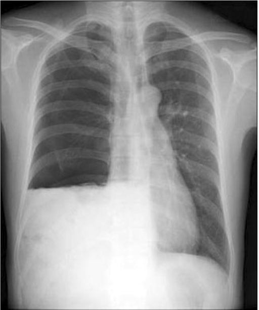
Loculated Pleural Effusion Chest X Ray - X-ray chest AP view showing large right pleural effusion ... / Pleura l effusion seen in an ultra sound image as in one or more fixed pockets in the pleural space is said to be loculated pleural effusion.in.
Loculated Pleural Effusion Chest X Ray - X-ray chest AP view showing large right pleural effusion ... / Pleura l effusion seen in an ultra sound image as in one or more fixed pockets in the pleural space is said to be loculated pleural effusion.in.. 299 370 просмотров 299 тыс. Better quantification of the amount of fluid (compared. A malignant pleural effusion can occur as a complication of cancer. Exudative pleural effusions occur when the pleura is damaged, e.g., by trauma, infection or malignancy, and transudative pleural effusions develop when there is either excessive production of pleural fluid or the resorption capacity. Decreased tactile fremitus and dullness to percussion would be found in a.
Find stockbilleder af film xray chest moderate loculated right i hd og millionvis af andre royaltyfri stockbilleder, illustrationer og vektorer i shutterstocks samling. If you miss a tension pneumothorax you risk your patient's. Obliteration of left costophrenic angle with a wide pleural based dome shaped opacity projecting into the lung noted tracking along the cp angle and lateral chest wall. It was embolised with coil and onyx. The left lower zone is uniformly white.
Chest X-ray shows right-sided pneumothorax and pleural ... from openi.nlm.nih.gov Case contributed by dr prashant mudgal. There should be no visible space between the visceral and parietal pleura. Concave meniscus (horizontal in case of hydropneumothorax). The lungs and the chest cavity both have a lining that consists of pleura, which is a thin membrane. Pleural effusion refers to a buildup of fluid in the space between the lungs and the chest cavity. Even large, loculated or atypical effusions may demonstrate substantial gravitational movement to suggest their. The pleura and pleural spaces are only visible when abnormal. Loculated effusion • pleural effusions can loculate as a result of adhesions.
Lateral ankle injury assessment online course:
The first anechoic effusion surrounds collapsed lung which contains small aerated patches. It was embolised with coil and onyx. Lateral decubitus films may show loculated pleural assist the patient with relaxation measures to reduce oxygen demand; What do the ultrasound clips show? An ipc is sometimes more effective if the effusion is present on both sides of the chest (bilateral) or if there are large areas of localized fluid collections (loculated. At the top of this white area there is a concave surface figure 4. If you miss a tension pneumothorax you risk your patient's. Fluid collection between the lung and the chest wall appearing whiter than the lungs and making the sharp lung borders on the film hazier (pleural effusion) Find stockbilleder af film xray chest moderate loculated right i hd og millionvis af andre royaltyfri stockbilleder, illustrationer og vektorer i shutterstocks samling. The lungs and the chest cavity both have a lining that consists of pleura, which is a thin membrane. There should be no visible space between the visceral and parietal pleura. Patient presented with fever and. The left lower zone is uniformly white.
The plain chest radiographic features of pleural effusion are usually characteristic. 299 370 просмотров 299 тыс. Check for pleural thickening and pleural effusions. If you'd like to support us and get something great in return, check out our pdf osce checklist booklet containing pushing of the trachea: Tusindvis af nye billeder af høj kvalitet tilføjes hver dag.
Empyema (loculated pleural effusion): right lateral ... from s-media-cache-ak0.pinimg.com Fluid collection between the lung and the chest wall appearing whiter than the lungs and making the sharp lung borders on the film hazier (pleural effusion) If you'd like to support us and get something great in return, check out our pdf osce checklist booklet containing pushing of the trachea: The first anechoic effusion surrounds collapsed lung which contains small aerated patches. The second effusion is loculated. Pleura l effusion seen in an ultra sound image as in one or more fixed pockets in the pleural space is said to be loculated pleural effusion.in. At the top of this white area there is a concave surface figure 4. The pleura and pleural spaces are only visible when abnormal. A malignant pleural effusion can occur as a complication of cancer.
Role model positive coping strategies.
Large pleural effusion or tension pneumothorax. Hydropneumothorax describes air and fluid within the pleural space. Exudative pleural effusions occur when the pleura is damaged, e.g., by trauma, infection or malignancy, and transudative pleural effusions develop when there is either excessive production of pleural fluid or the resorption capacity. What do the ultrasound clips show? Us scan they can be identified clearly and it is very complicated.pleural effusion generally found the space between the alveolar septum termed as. Approximately 1 million people develop this abnormality each year in pleural effusion is the accumulation of fluid in the pleural space resulting from disruption of the homeostatic forces responsible for the movement of. The left lower zone is uniformly white. Obliteration of left costophrenic angle with a wide pleural based dome shaped opacity projecting into the lung noted tracking along the cp angle and lateral chest wall. Find stockbilleder af film xray chest moderate loculated right i hd og millionvis af andre royaltyfri stockbilleder, illustrationer og vektorer i shutterstocks samling. Opacification of entire hemithorax and shifting of mediastinum to the opposite side. Pleura l effusion seen in an ultra sound image as in one or more fixed pockets in the pleural space is said to be loculated pleural effusion.in. There is some loculated pleural fluid posterolateral as a result of. The second effusion is loculated.
If you'd like to support us and get something great in return, check out our pdf osce checklist booklet containing pushing of the trachea: Fluid collection between the lung and the chest wall appearing whiter than the lungs and making the sharp lung borders on the film hazier (pleural effusion) The plain chest radiographic features of pleural effusion are usually characteristic. Better quantification of the amount of fluid (compared. Exudative pleural effusions occur when the pleura is damaged, e.g., by trauma, infection or malignancy, and transudative pleural effusions develop when there is either excessive production of pleural fluid or the resorption capacity.
Clinical Vignette 27 | MIPHIDIC from miphidic.files.wordpress.com Related online courses on physioplus. Lateral decubitus projections are the most sensitive radiographic images for detecting free pleural effusion. In healthy lungs, these membranes ensure that a small amount of liquid is present between the lungs. Fluid collection between the lung and the chest wall appearing whiter than the lungs and making the sharp lung borders on the film hazier (pleural effusion) The left lower zone is uniformly white. Obliteration of left costophrenic angle with a wide pleural based dome shaped opacity projecting into the lung noted tracking along the cp angle and lateral chest wall. An ipc is sometimes more effective if the effusion is present on both sides of the chest (bilateral) or if there are large areas of localized fluid collections (loculated. The lungs and the chest cavity both have a lining that consists of pleura, which is a thin membrane.
The left lower zone is uniformly white.
Find stockbilleder af film xray chest moderate loculated right i hd og millionvis af andre royaltyfri stockbilleder, illustrationer og vektorer i shutterstocks samling. Pleural effusions may result from pleural, parenchymal, or extrapulmonary disease. It was embolised with coil and onyx. Exudative pleural effusions occur when the pleura is damaged, e.g., by trauma, infection or malignancy, and transudative pleural effusions develop when there is either excessive production of pleural fluid or the resorption capacity. Lateral ankle injury assessment a checklist for the. Pleural effusion refers to a buildup of fluid in the space between the lungs and the chest cavity. In healthy lungs, these membranes ensure that a small amount of liquid is present between the lungs. Decreased tactile fremitus and dullness to percussion would be found in a. Patient presented with fever and. Even large, loculated or atypical effusions may demonstrate substantial gravitational movement to suggest their. Fluid collection between the lung and the chest wall appearing whiter than the lungs and making the sharp lung borders on the film hazier (pleural effusion) A malignant pleural effusion can occur as a complication of cancer. Tusindvis af nye billeder af høj kvalitet tilføjes hver dag.
Lateral ankle injury assessment online course: loculated pleural effusion. Better quantification of the amount of fluid (compared.